Amoebiasis: definition, symptoms and diagnosis
Amoebiasis
Amebiasis is a digestive disease caused by a single-celled parasite or protozoa in scientific terms, which is caused by Entamoeba. This parasite has various species and the most important species that can cause diseases and digestive problems for humans is called Entamoeba histolytica (E. histolytica). This parasite can cause infection in the digestive tract and infections outside the digestive tract. Other species of this single-celled parasite have also been identified, which do not cause any special disease or problem for humans. These types include Moshkovskiy, Dispar and Bangladeshi.
According to the statistics announced by the Center for Disease Control and Prevention (CDC), acute symptoms of this disease are observed in only about 10-20% of people with amebiasis, but the important thing is that even people without symptoms can spread cysts. new in the environment, spread the disease.
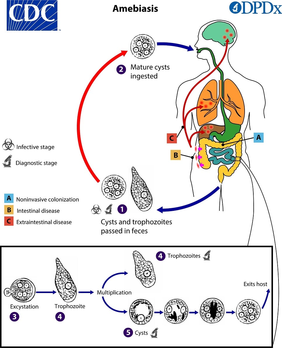
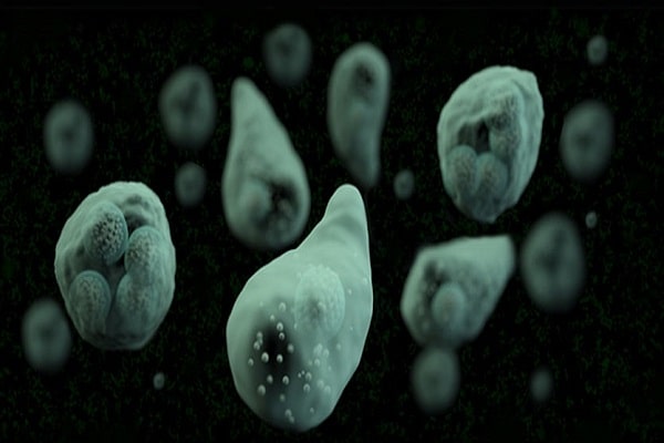
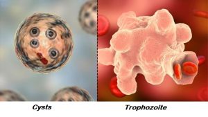
The aggressive and active form of this parasite called trophozoite. Its cyst are transmitted by feces. Mainly, the cyst is formed in the formed stool (hard) and its trophozoite is formed in the diarrhea stool (loose). Infection with this parasite occurs by swallowing its cyst through water or food or from hands contaminated with feces. The cysts are opened in the small intestine and the trophozoite is released and transferred to the large intestine. It is possible that the cysts remain in the lumen of the large intestine and cause the non-invasive form of this disease and the person continues to excrete the cyst from the intestine, or the trophozoites invade the intestinal wall and blood vessels and are detected in organs such as the liver, lungs, and brain. and reproduce.
Cysts can survive for a few days to a few weeks in the environment outside the body due to their protective wall and have the ability to cause disease if swallowed, but the trophozoite form is destroyed even if it is swallowed due to the acidic conditions of the stomach.
Who is at risk for amebiasis?
A wide geographic distribution has been defined for this disease. Mainly, humans are infected with this single-celled parasite by drinking water contaminated with human feces. Amoebiasis is common in tropical countries with underdeveloped sanitation. This disease is common in the Indian subcontinent, parts of Central and South America, Mexico and parts of Africa and relatively rare in the United States. People who are most at risk of developing amoebiasis include:
- People who have traveled to tropical areas where there is no good sanitation.
- Immigrants from tropical countries with unfavorable health conditions.
- People living in institutions with undeveloped sanitary conditions such as prisons.
- Men who have sex with other men.
- People with suppressed immune systems and other adverse health conditions.
In most patients, the infection is limited to the large intestine and is asymptomatic. Colitis or inflammation of the large intestine is caused by invasion of the wall of the large intestine. Dysentery or bloody diarrhea is usually the result of infection with this parasite. In case of chronic infection with Entamoebahistolytica, complications such as granuloma and peritonitis may occur due to the destruction of intestinal tissue. Liver abscess is one of the most important extraintestinal diseases caused by histolytica, which causes fever and pain in the upper right part of the abdomen. On the other hand, brain-pulmonary abscesses, vaginal lesions and necrotic lesions are also observed in the skin around the anus.
How is amoebic diarrhea diagnosed?
To diagnose amebiasis and rule out other possible infections, the attending physician may prescribe a variety of tests, from direct stool testing to specialized tests. In general, common tests to diagnose this parasitic disease include:
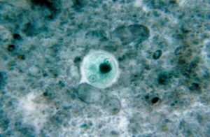
Direct examination of feces: in laboratory investigations, direct observation of feces based on the observation of red blood cells swallowed by Entamoeba histolytica, which is considered as a suitable finding to differentiate this pathogenic species from other amoeba species. Of course, the number and structural characteristics of the nucleus and inside the cell are guiding for an experienced parasitology technician. Trichrome and hematoxylin staining techniques are also used to identify protozoa more accurately.
Antigen test: The patient’s stool sample may be tested for Entamba histolytica antigen. This is done through a method called enzyme-linked immunosorbent assay (ELISA).
Molecular test (PCR): Molecular polymerase chain reaction (PCR) test, both conventional PCR and real-time PCR, may be performed to distinguish amoebiasis (DNA) from other infections. According to the World Health Organization, the use of PCR has been recommended as an effective tool for epidemiological studies and clinical diagnosis.
Among the various methods mentioned, microscopic observation (due to the lack of differentiation between different species of amoeba) and serology tests (due to various reasons, including the lack of differentiation of current infection from the past and cross-reactions), have not been effective and nowadays are used for correct and early diagnosis. , the development and application of new molecular methods have been emphasized with an emphasis on preventing indiscriminate and incorrect drug use.
Resources: